熒光 微球分析技術屬于化學材料發展結果,可用于細胞表面抗原的檢測、退行性神經病變示蹤物、吞噬功能的檢測、血流分析、敏感性診斷試劑等,本文介紹了熒光微球分析技術以及熒光微球吞噬實驗的操作步驟。
熒光微球分析 技術簡介
熒光微球分析技術是近年來化學材料科學活躍發展 的產物,各種大小(0.2~10μm)可產生熒光和色彩的人工微球應運而生,目前各種材料人工合成、多種顏色和規格的熒光微球有2大類,一種是表面不帶修飾基團的微球,另一種是攜帶各種化學修飾基團,包括羧基化修飾、氨基修飾、巰基或醛巰基修飾等的微球。
應用
這些熒光微球正廣泛應用于生命科學研究的多個領域中,如:
(1)細胞表面抗原的檢測,包括CD4/CD8表面抗原的絕對計數;細胞表面低豐度表達受體的分析;骨髓移植受體內供體紅細胞的檢測;白色念珠菌抗原檢測等。
(2)退行性神經病變示蹤物,熒光微球具有無毒、與神經細胞結合時間長以及受注射部位影響極小的特點。
(3)吞噬功能的檢測,0.6~2.0μm大小的熒光微球適合于這一類的研究,如分析大鼠中性粒細胞、人橫紋導管細胞、小鼠腹膜巨噬細胞、人多核白細胞的吞噬功能或不同調理素調理作用對吞噬功能的影響等。
(4)血流分析,10-15 μm大小7種顏色的熒光可供研究組織中局部血流情況,如腫瘤脈管血流速率、視網膜和脈絡膜循環、肺泡微管的功能直徑定位等。
(5)敏感性診斷試劑,如替代一些已開展應用的微球診斷試驗:膠乳凝集試驗、微球捕獲ELASA、雙位點夾心法等,它較傳統比色方法更為靈敏;另有新近誕生的流式微球分析技術(Cytometric Bead Array CBA)通過將不同熒光強度的聚苯乙烯微球包被上多種高特異性的單克隆抗體,在微球“三明治”平臺上進行可溶性蛋白的檢測,由此而使得1份微量標本可同時1次定量分析多種可溶性蛋白,大大節約了樣本量和操作時間,靈敏度也較傳統的ELISA方法更高,重復性好。
(6)質量控制,2~10 μm直徑,大小均一,表面定量吸附不同熒光素的樹脂顆粒可用于流式細胞儀的質控,在樣品測試前調整儀器對測試信號的放大系數,以保證各次實驗結果具有高度的重復性,這類微球標準品按其應用被分為以下2類:即定量熒光標準微球和準值熒光標準微球。
熒光微球吞噬實驗的Protocol
---Materials
*Fluorescence labeled latex beads (1um diameter), 2.5% aqueous suspension
* 3% BSA containing 25 mM Na2HPO4, pH 6.0
* 0,3%(w/v) azide
* Culture medium containing 5% FBS
* Distilled water, PBS
* Bath sonication, 6(12)-well plates
---Cell culture and treatment
1, Inoculate plates with 7,0?104 cells/cm2 per well. Incubate at 37℃,5% CO2 for 24 hr, best until 50-70% confluence is reached
2, Remove culture medium and expose the cells to test material. Incubate at 37℃,5% CO2 for 24 hr.
---Preparation of coated latex beads
1, Wash latex beads with distilled water and pellet at 10,000g for 8 min at RT.
2, Resuspend latex beads in 3% BSA containing 25 mM Na3PO4 (pH 6.0) and incubate at RT for 15 min with bath sonication.
* Coating beads in BSA insures beads remain in a monodisperse state.
3, Wash the beads once with culture medium containing 5% FBS.
4 ,Resuspend the beads in culture medium at concentration 2.0%.This is beads stock. Stored in darkness at 4℃.
---Assay
1, Controls and samples: Intact control (No staining) 1 well
-Negative control (azide treated) 1 well
-Normal control 1 well
-sample 5 wells
* In order to differentiate between phagocytosed beads and beads nonspecifically adhere to the cell surface, control cells are exposed to 0,3%(w/v) azide for 10 min prior to the addition of coated beads. This treatment compromises microglial energetic processes and few beads were internalized as observed by fluorescent microscopy. Mean fluorescence of azide-treated microglia was used as the negative control and was subtracted from values obtained in experimental samples.
2, For experiments using 6 well-plates, 15 μl beads stock in 1 ml culture medium is applied to each well. Votex the beads stock well and take out 105 μl and add into 7 ml culture medium. Bath sonificate for 10 min at RT in darkness. This is beads working solution.
For experiments using 12 well-plates, 6 μl beads stock in 0.4 ml culture medium is applied to each well.
3, Wash each well twice with PBS and replace with beads working solution, 1 ml/well for 6-well plate, 0.4 ml/well for 12 well-plate. Incubate in the dark at 37 ℃ for 80-120 min.
4, Remove beads working solution and wash 3 times with PBS to remove excess beads.
5, Lift the cells by scrapping or trypsinization and wash the cells wish PBS.
6, Stain with PI (4ug/ml final concentration) and run for FACS.
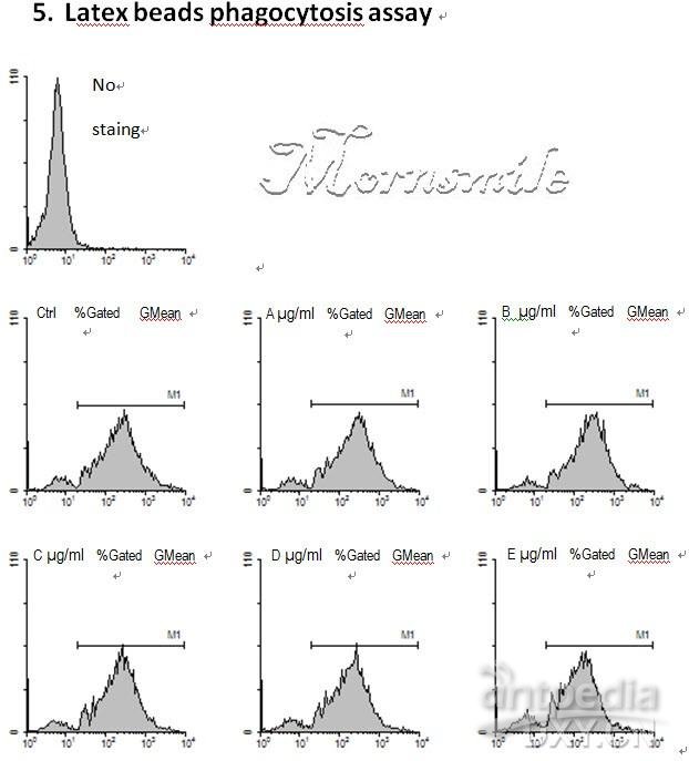
此3D圖是我的結果。
所用細胞并非專職吞噬細胞(astrocyte)。
隨處理劑量增大,可見熒光細胞的比例以及熒光強度逐漸降低。
最下面灰色的細胞群是無熒光對照。

下圖是Dot plot
圖形特點:感嘆號

下圖是專職吞噬細胞(microglia)對熒光微球的吞噬。
可見所給處理對吞噬功能無明顯影響。
Histogram
很多同學都在找熒光微球體吞噬實驗操作步驟,看到這篇資料非常不錯,很清楚的了解微球體吞噬實驗protocol,在這里分享給大家,希望對剛做熒光微球體吞噬實驗的同學有幫助。